MAGNETIC STIMULATION IN APHASIA
1.- INTRODUCTION
After suffering a stroke, from the acute post-lesional phase, a reorganization of neural networks that have not been affected is generated, so that the healthy neurons can “learn” functions from the damaged neurons and then replace them, leading to neuronal plasticity or neuroplasticity. Neuroplasticity or neural plasticity is the ability to auto-reorganise nerve tissue.
This phenomenon (neuroplasticity) is determined by genetic factors, patient age, degree of dependency prior to the damaging event, earliness with which neurorehabilitation, social and family support, intercurrent complications, as well as the location, intensity, nature and extent of the brain injury begin.
Most symptoms after stroke are not only due to the injury itself, but due to the hyperactivity registered in the hemisphere intact to the injured hemisphere, which is inhibited.
On the other hand, TMS (Transcranial Magnetic Stimulation) accelerates neuroplasticity mechanisms, quickly reorganizing brain connections, which leads to greater efficiency of neural networks in the affected area.
Low frequency (≤ 1 Hz) repetitive TMS (rTMS) applied to the healthy hemisphere reduces diffuse cortical activation, after a stroke, in the primary and secondary motor areas of both brain hemispheres, activating the injured cortical area that had been inhibited and encouraging its excitability and motor recovery. However, low frequency rTMS (≥ 5 Hz) increases cortical excitability and can be applied to produce a neural stimulation of the cortex of the injured hemisphere. Meaning it accelerates neuroplasticity mechanisms, quickly reorganizing brain connections, which leads to greater efficiency of neural networks in the affected area.

TMS has come to be considered a therapeutic reality for neurodegenerative, psychiatric, neurological diseases and other clinical specialties conferring neuroprotective effects that impact positively on the modulation of neuroplasticity, helping the brain in its ability to renew and/or reconnect neural circuits and thus acquire new skills.
TMS in stroke can also be used as a regenerative therapy technique.
With regard to its therapeutic effects in stroke patients, TMS can be focused towards enhancing neuroplasticity, and thus towards each of the symptoms associated with stroke (motor recovery, language and swallowing disorders, depression and perceptual and cognitive impairments).
The basis of this neurorehabilitation therapy is based on the fact that the brain is a dynamic entity adaptable to both internal and external environmental changes.
TMS is a technique that allows us to act positively on these neural changes in a safe and non-invasive, provided that it is implemented by an experienced team: The intensity of the electromagnetic pulse in the implementation of the TMS is an individual and specific measure for each patient, therefore guidelines and protocols have to be followed, given that there is variability depending on the medical and therapeutic equipment used to apply it.

2. – WHAT IS APHASIA?
Aphasia is a disorder that affects the ability to express and/or understand language, usually following an injury in the perisylvian region of the left cerebral hemisphere.
In short, aphasia is an impairment acquired in the ability to emit and/or understand language, both oral and written, as well as gestural.
Language is the vehicle of thought. Aphasia disorders almost always involve other functions of the written language (dysgraphia) and reading (alexia).
The aphasic patients clinic will act according to the location and size of the brain injury, as well as the cerebral capacity of healthy neurons to assume the functions of the injured ones (what we call neural plasticity or neuroplasticity).
Sometimes, after the harmful event, the organizational changes in the interneuronal brain activity of the affected area and the surrounding healthy regions can recover language capacity. Therefore when patients suffering from aphasia go beyond the acute period of convalescence and are stabilized, they should receive speech therapy in order to help achieve this objective.
The cause of aphasia is varied: a stroke or cerebral infarction (this is the most common cause), a head injury, an infection of the brain, a brain tumour, dementia, etc.
The right and left brain hemispheres have different functions with regard to language, the left is more specialized in the lexical and syntactic aspects, and right in the prosodic aspects or “emotional” aspects of language.

3- WHAT IS TRANSCRANIAL MAGNETIC STIMULATION (TMS)?

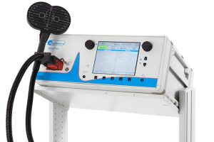
TMS is a non-invasive form of stimulation of the cerebral cortex, and represents a technical tool that extends the range of possibilities for study and research in the field of neuroscience, as well as in the treatment of various diseases and neuropsychiatric disorders. It allows safe, painless and non-invasive stimulation of the nervous tissue (cerebral cortex, spinal cord, central motor pathways and peripheral nerves) as well as the controlled regulation of brain activity.
Fundamentals:
TMS is based on the principle of electromagnetic induction discovered by Michael Faraday in 1831. An electric current passes through a coil of copper wire encapsulated in a plastic housing, located on the patient’s head. When a pulse of current passes through the stimulation coil, a magnetic field is generated which passes through the scalp and cranial vault undiminished. This magnetic field which is variable over time induces an electric current in the neuronal tissue of the brain, whose volume depends on the shape, size, type and orientation of the coil, the strength (intensity) of the magnetic field and the frequency and duration of the magnetic pulses produced. Thus, TMS might be considered a form of non-invasive electrodeless electrical stimulation by electromagnetic induction.

This electric current acts on brain cells (neurons) inhibiting or stimulating their effects.
From the therapeutic perspective, there are a lot of studies that show that transcranial magnetic stimulation is effective and can also be considered safe, provided that it is used by a qualified medical team and safety guidelines are met.
4- CURRENT THERAPEUTIC APPLICATIONS AND USE OF THE TMS:
Since TMS is a non-invasive, well tolerated technique, with few contraindications, it has become a cutting edge therapy, used to treat various disorders, both psychiatric and neurological (especially in patients with cerebrovascular pathology), and is approved by the Food and Drug Administration of the United States as a treatment of choice when the patient experiences a major depression refractory with conventional drug treatment.

Its applications are numerous and increasingly broad thanks to research that is emerging every year:
Aphasia: Aphasia, in its various forms, is a common consequence of stroke, especially in the left hemisphere, characterized by impairments in speech, comprehension, reading and writing. Treatment with TMS is more efficient, based on published scientific literature, in patients with motor aphasia (not fluent) or in global forms of aphasia with motor predominance.
Several studies have helped to confirm that stimulation alone improves language disorders in the identification of images, such as in spontaneous speech, and repeatition, nomination and comprehension tests.
Oropharyngeal dysphagia: Although its incidence is 50% in stroke patients, oropharyngeal dysphagia is underestimated and underdiagnosed, constituting a cause of malnutrition and aspiration pneumonia, which increases the mortality rate in these patients (20-30% of post-stroke deaths).
Oropharyngeal dysphagia produces two types of complications: impairments in swallowing efficiency (causing malnutrition or dehydration) and insecurity swallowing (which can produce aspiration pneumonia). Dysphagia after a stroke is a result of the damage to the dominant motor cortex.

5.- OUTLINE OF THE TREATMENT
Before starting the first rTMS session, a medical visit is carried out to verify that the patient presents no contraindication and the patient is able to participate in the treatment. A speech therapy assessment is also performed before and after treatment, and subsequent follow-up visits are made to assess the response to this type of combined neurorehabilitation therapy.
APHASIA TREATMENT PLAN: The treatment protocol for aphasia that takes place at the San Vicente Clinic is based on the protocol developed by the Berenson-Allen Center for Noninvasive Brain Stimulation (CNBS) at Beth Israel Deaconess Medical Center and Harvard Medical School, whose scientific support is based on research work carried out mainly by Margaret Naeser and her colleagues.
It consists of 10 sessions of rTMS (a daily session for 10 working days of two weeks) of 20 continual minutes of intensive speech therapy (approximately 2 hours daily).
DYSPHAGIA TREATMENT PLAN: The application of the stimulation is applied 10 minutes a day for 2 weeks, on the contralesional motor cortex, having demonstrated the improvement in swallowing and decreased risk of aspiration after treatment.
6.- SIDE EFFECTS
TMS is a safe technique provided that safety guidelines are followed. Some patients undergoing this cortical stimulation may experience side effects after application, which might be considered mild and transient, such as cephalic or cervical pain; and in the rare situation of persistence, this is mitigated by taking common painkillers.
Moreover, the risk of seizures during TMS is very low and it has not been shown that TMS increases the risk of seizures in patients with controlled epilepsy, once the stimulation session has ended.
7.- CONTRAINDICATIONS
The main relative contraindications that TMS has are: women who are pregnant and children under six.
The following is a list of patients that have absolute contraindications: those with pacemakers, deep brain stimulation electrodes, personal electronic devices (drug infusion pumps) or intracranial metallic elements (metal plates, wires, screws, heart valves or ventriculoperitoneal bypass, cochlear implants, etc.). Nor should treatment be performed on patients with uncontrolled epilepsy. Before starting treatment a doctor will evaluate each case individually, to rule out any contraindications.

8.- CONCLUSIONS
- TMS has proven to be a cutting-edge technical ally, safe and effective in treating the deficits that may arise after a stroke, as well as in terms of safety, it is innocuous to the patient. Likewise, TMS has proven to be especially valuable in helping to promote brain regeneration by means of the neuroplasticity mechanism.
- Excitatory and inhibitory electromagnetic pulses applied in the ipsilateral or contralateral cerebral hemisphere to the lesion, as well as the transcallosal area in order to modulate communication between the two brain hemispheres (depending on the desired effect), offer us the possibility of optimizing functional brain activity by inducing changes in interhemispheric connectivity, as well as achieving recovery of the damaged brain area in less time.
- The various studies conducted in the field of TMS have confirmed the improvement of motor disorders, aphasia, spasticity, oropharyngeal dysphagia and perceptual and cognitive difficulties that occur in patients with a stroke.

9.- BIBLIOGRAPHY
- Ferro B, Aragmende D. Importancia del logopeda para los pacientes con trastornos del lenguaje y de la deglución. En: Castillo Sánchez J, Jiménez Martín I. Reeducación funcional tras un ictus. Barcelona: Elsevier España, S.L.U.; 2015. p. 161-81.
- Figueroa J, Villamayor B, Antelo A. Rehabilitación del ictus cerebral: evaluación, pronóstico y tratamiento. En: Castillo Sánchez J, Jiménez Martín I. Reeducación funcional tras un ictus. Barcelona: Elsevier España, S.L.U.; 2015. p. 89-104.
- Campos F, Sobrino T, Sánchez JC. Estrategias neuroprotectoras en el ictus isquémico. En: Castillo Sánchez J, Jiménez Martín I. Reeducación funcional tras un ictus. Barcelona: Elsevier España, S.L.U.; 2015. p. 63-73.
- Sobrino T, Campos F, Sánchez JC. Nuevas líneas de futuro: la terapia celular. En: Castillo Sánchez J, Jiménez Martín I. Reeducación funcional tras un ictus. Barcelona: Elsevier España, S.L.U.; 2015. p. 75-85.
- Raffin E, Siebner HR. Transcranial brain stimulation to promote functional recovery after stroke. Curr Opin Neurol. 2014; 27: 54-60.
- Escribano MB, Túnez I. Estimulación magnética transcraneal como nueva estrategia terapéutica en el ictus. En: Castillo Sánchez J, Jiménez Martín I. Reeducación funcional tras un ictus. Barcelona: Elsevier España, S.L.U.; 2015. p. 121-33.
- Edwardson MA, Lucas TH, Carey JR, Fetz EE. New modalities of brain stimulation for stroke rehabilitation. Exp Brain Res. 2013; 224: 335-58.
- Lefaucheur JP, André-Obadia N, Antal A, Ayache SS, Baeken C, Benninger DH, et al. Evidence-based guidelines on the therapeutic use of repetitive transcranial magnetic stimulation (rTMS). Clin Neurophysiol. 2014; 125: 2150-206.
- The Brain and Behavior. Language. In: Bear MF, Connors BW, Paradiso MA, editors. Neuroscience: exploring the brain. 4th ed. Philadelphia: Wolters Kluwer; 2015. p. 685-718.
- Somme J, Zarranz JJ. Trastornos de las funciones cerebrales superiores. Alteraciones del lenguaje y del habla. En: Zarranz JJ, editor. Neurología. 5ª ed. Barcelona: Elsevier España, S.L.; 2013. p. 170-6.
- Mayo Clinic Staff. Diseases and Conditions. Aphasia. Basics. Causes. Available at: http://www.mayoclinic.org/diseases-conditions/aphasia/basics/causes/con-20027061 (updated March 21, 2015; accessed August 03, 2015).
- Ardila A. Daño cerebral en la afasia. En: Ardila A, editor. Las afasias. Miami: Ardila A. 2006. p. 26-47.
- Pascual-Leone A, Tormos-Muñoz JM. Estimulación magnética transcraneal: fundamentos y potencial de la modulación de redes neuronales específicas. Rev Neurol. 2008; 46: S3-10.
- Verdugo-Díaz L, Drucker-Colin R. Campos magnéticos: usos en la biología y la medicina. En: Túnez Fiñana I, Pascual Leone A. Estimulación magnética transcraneal y neuromodulación. Presente y futuro en neurociencias. Barcelona: Elsevier España, S.L.; 2014. p. 1-19.
- Medina FJ, Pascual A, Túnez I. Mecanismos de acción en la estimulación magnética transcraneal En: Túnez Fiñana I, Pascual Leone A. Estimulación magnética transcraneal y neuromodulación. Presente y futuro en neurociencias. Barcelona: Elsevier España, S.L.; 2014. p. 21-30.
- Barker AT, Jalinous R, Freeston IL. Non-invasive magnetic stimulation of the human motor cortex. 1985; 1: 1106-7.
- Barker AT. The history and basic principles of magnetic nerve stimulation. In: Pascual-Leone A, Davey N, Rothwell J, Wasserman E, Puri B, editors. Handbook of transcranial magnetic stimulation. London: Arnold; 2002. p. 3-17.
- Kobayashi M1, Pascual-Leone A. Transcranial magnetic stimulation in neurology. Lancet Neurol. 2003; 2: 145-56.
- Rossi S, Hallett M, Rossini PM, Pascual-Leone A; Safety of TMS Consensus Group. Safety, ethical considerations, and application guidelines for the use of transcranial magnetic stimulation in clinical practice and research. Clin Neurophysiol. 2009; 120; 2008-39.
- Emara TH, Moustafa RR, Elnahas NM, Elganzoury AM, Abdo TA, Mohamed SA, et al. Repetitive transcranial magnetic stimulation at 1Hz and 5Hz produces sustained improvement in motor function and disability after ischaemic stroke. Eur J Neurol. 2010; 17: 1203-9.
- Bayón M. Estimulación magnética transcraneal en la rehabilitación del ictus. Rehabilitación (Madr). 2011; 45: 261-7.
- Kakuda W, Abo M, Momosaki R, Morooka A. Therapeutic application of 6-Hz-primed low-frequency rTMS combined with intensive speech therapy for post-stroke aphasia. Brain Inj. 2011; 25: 1242-8.
- Corti M, Patten C, Triggs W. Repetitive transcranial magnetic stimulation of motor cortex after stroke: a focused review. Am J Phys Med Rehabil. 2012; 91: 254-70.
- Wassermann EM, Zimmermann T. Transcranial magnetic brain stimulation: therapeutic promises and scientific gaps. Pharmacol Ther. 2012; 133: 98-107.
- Chervyakov AV, Chernyavsky AY, Sinitsyn DO, Piradov MA. Possible Mechanisms Underlying the Therapeutic Effects of Transcranial Magnetic Stimulation Front Hum Neurosci. 2015; 9: 303.
- Camprodon JA. Integración de la estimulación magnética transcraneal con técnicas de neuroimagen. En: Túnez Fiñana I, Pascual Leone A. Estimulación magnética transcraneal y neuromodulación. Presente y futuro en neurociencias. Barcelona: Elsevier España, S.L.; 2014. p. 55-66.
- Espinosa N, Arias P, Cudeiro J. La estimulación magnética transcraneal como instrumento para el estudio del sistema visual. En: Túnez Fiñana I, Pascual Leone A. Estimulación magnética transcraneal y neuromodulación. Presente y futuro en neurociencias. Barcelona: Elsevier España, S.L.; 2014. p. 67-78.
- García-Toro M, Gili M, Roca M. Estimulación magnética transcraneal en psiquiatría. En: Túnez Fiñana I, Pascual Leone A. Estimulación magnética transcraneal y neuromodulación. Presente y futuro en neurociencias. Barcelona: Elsevier España, S.L.; 2014. p. 79-86.
- Valls-Solé J. La estimulación magnética en el estudio de lesiones medulares. En: Túnez Fiñana I, Pascual Leone A. Estimulación magnética transcraneal y neuromodulación. Presente y futuro en neurociencias. Barcelona: Elsevier España, S.L.; 2014. p. 87-100.
- Mondragón H, Alonso M. Aplicación de la estimulación magnética transcraneal en la patología cerebrovascular. En: Túnez Fiñana I, Pascual Leone A. Estimulación magnética transcraneal y neuromodulación. Presente y futuro en neurociencias. Barcelona: Elsevier España, S.L.; 2014. p. 101-14.
- Tasset I, Agüera E, Sánchez F. Realidad actual de la aplicación de EMT a los trastornos neurodegenerativos y neuropsiquiátricos. En: Túnez Fiñana I, Pascual Leone A. Estimulación magnética transcraneal y neuromodulación. Presente y futuro en neurociencias. Barcelona: Elsevier España, S.L.; 2014. p. 115-25.
- Bartrés-Faz D, Peña-Gómez C. Estimulación cerebral no invasiva, redes neuronales y diferencias individuales moduladoras. En: Túnez Fiñana I, Pascual Leone A. Estimulación magnética transcraneal y neuromodulación. Presente y futuro en neurociencias. Barcelona: Elsevier España, S.L.; 2014. p. 41-54.
- Carrera E, Tononi G. Diaschisis: past, present, future. Brain. 2014; 137: 2408-22.
- Liew SL, Santarnecchi E, Buch ER, Cohen LG. Non-invasive brain stimulation in neurorehabilitation: local and distant effects for motor recovery. Front Hum Neurosci. 2014; 8: 378.
- Cunningham DA, Machado A, Janini D, Varnerin N, Bonnett C, Yue G, et al. Assessment of inter-hemispheric imbalance using imaging and noninvasive brain stimulation in patients with chronic stroke. Arch Phys Med Rehabil. 2015; 96: S94-103.
- Di Pino G, Pellegrino G, Assenza G, Capone F, Ferreri F, Formica D, et al. Modulation of brain plasticity in stroke: a novel model for neurorehabilitation. Nat Rev Neurol. 2014; 10: 597-608.
- Simonetta-Moreau M. Non-invasive brain stimulation (NIBS) and motor recovery after stroke. Ann Phys Rehabil Med. 2014; 57: 530-42.
- Malcolm MP, Vaughn HN, Greene DP. Inhibitory and excitatory motor cortex dysfunction persists in the chronic poststroke recovery phase. J Clin Neurophysiol. 2015; 32: 251-6.
- Karabanov A, Ziemann U, Hamada M, George MS, Quartarone A, Classen J, et al. Consensus Paper: Probing Homeostatic Plasticity of Human Cortex With Non-invasive Transcranial Brain Stimulation. Brain Stimul. 2015; 8: 442-54.
- Cassidy JM, Chu H, Anderson DC, Krach LE, Snow L, Kimberley TJ, et al. A Comparison of Primed Low-frequency Repetitive Transcranial Magnetic Stimulation Treatments in Chronic Stroke. Brain Stimul. 2015 Jun 22. pii: S1935-861X(15)01009-8. doi: 10.1016/j.brs.2015.06.007 [Epub ahead of print].
- Thiel A, Black SE, Rochon EA, Lanthier S, Hartmann A, Chen JL, et al. Non-invasive repeated therapeutic stimulation for aphasia recovery: a multilingual, multicenter aphasia trial. J Stroke Cerebrovasc Dis. 2015; 24: 751-8.
- Demirtas-Tatlidede A, Alonso-Alonso M, Shetty RP, Ronen I, Pascual-Leone A, Fregni F. Long-term effects of contralesional rTMS in severe stroke: safety, cortical excitability, and relationship with transcallosal motor fibers. NeuroRehabilitation. 2015; 36: 51-9.
- Yoon TH, Han SJ, Yoon TS, Kim JS, Yi TI. Therapeutic effect of repetitive magnetic stimulation combined with speech and language therapy in post-stroke non-fluent aphasia. NeuroRehabilitation. 2015; 36: 107-14.
- Hosomi K, Seymour B, Saitoh Y. Modulating the pain network–neurostimulation for central poststroke pain. Nat Rev Neurol. 2015; 11: 290-9.
- Fuentes B, Gállego J, Gil-Nuñez A, Morales A, Purroy F, Roquer J, et al. Guidelines for the preventive treatment of ischaemic stroke and TIA (I). Update on risk factors and life style. Neurología. 2012; 27: 560-74.
- Blanco M. Aspectos demográficos y epidemiológicos del ictus. En: Castillo Sánchez J, Jiménez Martín I. Reeducación funcional tras un ictus. Barcelona: Elsevier España, S.L.U.; 2015. p. 11-20.
- Kim YH, You SH, Ko MH, Park JW, Lee KH, Jang SH, et al. Repetitive transcranial magnetic stimulation-induced corticomotor excitability and associated motor skill acquisition in chronic stroke. Stroke. 2006; 37: 1471-6.
- Hallett M. Transcranial magnetic stimulation: a primer. Neuron. 2007; 55: 187-99.
- Malcolm MP, Triggs WJ, Light KE, Gonzalez Rothi LJ, Wu S, Reid K, et al. Repetitive transcranial magnetic stimulation as an adjunct to constraint-induced therapy: an exploratory randomized controlled trial. Am J Phys Med Rehabil. 2007; 86: 707-15.
- Ameli M, Grefkes C, Kemper F, Riegg FP, Rehme AK, Karbe H, et al. Differential effects of high-frequency repetitive transcranial magnetic stimulation over ipsilesional primary motor cortex in cortical and subcortical middle cerebral artery stroke. Ann Neurol. 2009; 66: 298-309.
- Takeuchi N, Chuma T, Matsuo Y, Watanabe I, Ikoma KS. Repetitive transcranial magnetic stimulation of contralesional primary motor cortex improves hand function after stroke. Stroke. 2005; 36: 2681-6.
- Fregni F, Boggio PS, Valle AC, Rocha RR, Duarte J, Ferreira MJ, et al. A sham-controlled trial of a 5-day course of repetitive transcranial magnetic stimulation of the unaffected hemisphere in stroke patients. Stroke. 2006; 37: 2115-22.
- Di Lazzaro V, Profice P, Pilato F, Capone F, Ranieri F, Pasqualetti P, et al. Motor cortex plasticity predicts recovery in acute stroke. Cereb Cortex. 2010; 20: 1523-8.
- Talelli P, Greenwood RJ, Rothwell JC. Exploring Theta burst stimulation as an intervention to improve motor recovery in chronic stroke. Clin Neurophysiol. 2007; 118: 333-42.
- Huang YZ, Rothwell JC, Edwards MJ, Chen RS. Effect of physiological activity on an NMDA-dependent form of cortical plasticity in human. Cereb Cortex. 2008; 18: 563-70.
- Ackerley SJ, Stinear CM, Barber PA, Byblow WD. Combining theta burst stimulation with training after subcortical stroke. Stroke. 2010; 41: 1568-72.
- Talelli P, Wallace A, Dileone M, Hoad D, Cheeran B, Oliver R, et al. Theta burst stimulation in the rehabilitation of the upper Limb: a semirandomized, placebo-controlled trial in chronic stroke patients. Neurorehabil Neural Repair. 2012; 26: 976-87.
- Meehan SK, Dao E, Linsdell MA, Boyd LA. Continuous theta burst stimulation over the contralesional sensory and motor cortex enhances motor learning post-stroke. Neurosci Lett. 2011; 500: 26-30.
- Martin PI, Naeser MA, Theoret H, Tormos JM, Nicholas M, Kurland J, et al. Transcranial magnetic stimulation as a complementary treatment for aphasia. Semin Speech Lang. 2004; 25: 181-91.
- Naeser MA, Martin PI, Nicholas M, Baker EH, Seekins H, Kobayashi M, et al. Improved picture naming in chronic aphasia after TMS to part of right Broca’s area: an open-protocol study. Brain Lang 2005; 93: 95-105.
- Barwood CH, Murdoch BE, Whelan BM, Lloyd D, Riek S, O’Sullivan JD, et al. Improved language performance subsequent to low-frequency rTMS in patients with chronic non-fluent aphasia poststroke. Eur J Neurol. 2011; 18: 935-43.
- Naeser MA, Martin PI, Lundgren K, Klein R, Kaplan J, Treglia E, et al. Improved language in a chronic nonfluent aphasia patient after treatment with CPAP and TMS. Cogn Behav Neurol. 2010; 23: 29-38.
- Ren CL, Zhang GF, Xia N, Jin CH, Zhang XH, Hao JF, et al. Effect of low-frequency rTMS on aphasia in stroke patients: a meta-analysis of randomized controlled trials. PLoS One. 2014; 9: e102557.
- Otal B, Olma MC, Flöel A, Wellwood I. Inhibitory non-invasive brain stimulation to homologous language regions as an adjunct to speech and language therapy in post-stroke aphasia: a meta-analysis. Front Hum Neurosci. 2015; 9: 236.
- Li Y, Qu Y, Yuan M, Du T. Low-frequency repetitive transcranial magnetic stimulation for patients with aphasia after stoke: A meta-analysis. J Rehabil Med. 2015 Jul 15. doi: 10.2340/16501977-1988 [Epub ahead of print].
- Kakuda W, Abo M, Kaito N, Watanabe M, Senoo A. Functional MRI-based therapeutic rTMS strategy for aphasic stroke patients: a case series pilot study. Int J Neurosci. 2010; 120: 60-6.
- Dammekens E, Vanneste S, Ost J, De Ridder D. Neural correlates of high frequency repetitive transcranial magnetic stimulation improvement in post-stroke non-fluent aphasia: a case study. Neurocase. 2014; 20: 1-9.
- Rofes L, Vilardell N, Clavé P. Post-stroke dysphagia: progress at last. Neurogastroenterol Motil. 2013; 25: 278-82.
- Kedhr EM, Abo-Elfetoh N. Therapeutic role of rTMS on recovery of dysphagia in patients with lateral medullary syndrome and brainstem infarction. J Neurol Neurosurg Psychiatry. 2010; 81: 495-9.
- Momosaki R, Abo M, Kakuda W. Bilateral repetitive transcranial magnetic stimulation combined with intensive swallowing rehabilitation for chronic stroke Dysphagia: a case series study. Case Rep Neurol. 2014; 6: 60-7.
- Momosaki R, Abo M, Watanabe S, Kakuda W, Yamada N, Kinoshita S. Repetitive Peripheral Magnetic Stimulation With Intensive Swallowing Rehabilitation for Poststroke Dysphagia: An Open-Label Case Series. Neuromodulation. 2015 May 6. doi: 10.1111/ner.12308 [Epub ahead of print].
- Doeltgen SH, Bradnam LV, Young JA, Fong E. Transcranial non-invasive brain stimulation in swallowing rehabilitation following stroke–a review of the literature.Physiol Behav. 2015; 143: 1-9.
- Pisegna JM, Kaneoka A, Pearson WG Jr, Kumar S, Langmore SE. Effects of non-invasive brain stimulation on post-stroke dysphagia: A systematic review and meta-analysis of randomized controlled trials.Clin Neurophysiol. 2015 May 9. pii: S1388-2457(15)00309-0. doi: 10.1016/j.clinph.2015.04.069 [Epub ahead of print].
- Khedr EM, Abo-Elfetoh N, Rothwell JC. Treatment of post-stroke dysphagia with repetitive transcranial magnetic stimulation. Acta Neurol Scand. 2009; 119: 155-61.
- Verin E, Leroi AM. Poststroke dysphagia rehabilitation by repetitive transcranial magnetic stimulation: a noncontrolled pilot study. Dysphagia. 2009; 24: 204-10.
- Patel AT, Duncan PW, Lai SM. The relation between impairments and functional outcomes poststroke. Arch Phys Med Rehabil. 2000; 81: 1357-63.
- Lim JY, Kang EK, Paik NJ. Repetitive transcranial magnetic stimulation for hemispatial neglect in patients after stroke: an open-label pilot study. J Rehabil Med. 2010; 42: 447-52.
- Kim YK, Jung JH, Shin SHA comparison of the effects of repetitive transcranial magnetic stimulation (rTMS) by number of stimulation sessions on hemispatial neglect in chronic stroke patients. Exp Brain Res. 2015; 233: 283-9.
- Kim BR, Kim DY, Chun MH, Yi JH, Kwon JS. Effect of repetitive transcranial magnetic stimulation on cognition and mood in stroke patients: a double-blind, sham controlled trial. Am J Phys Med Rehabil. 2010; 89: 62-8.
- Xie Y, Zhang T, Chen AC. Repetitive Transcranial Magnetic Stimulation for the Recovery of Stroke Patients With Disturbance of Consciousness. Brain Stimul. 2015; 8: 674-5.
- Naeser MA, Martin PI, Baker EH, Hodge SM, Sczerzenie SE, Nicholas M, et al. Overt propositional speech in chronic nonfluent aphasia studied with the dynamic susceptibility contrast fMRI method. Neuroimage. 2004; 22: 29-41.
- Naeser MA, Martin PI, Nicholas M, Baker EH, Seekins H, Kobayashi M, et al. Improved picture naming in chronic aphasia after TMS to part of right Broca’s area: an open-protocol study. Brain Lang. 2005; 93: 95-105.
- Naeser MA, Martin PI, Nicholas M, Baker EH, Seekins H, Helm-Estabrooks N, et al. Improved naming after TMS treatments in a chronic, global aphasia patient–case report. Neurocase. 2005; 11: 182-93.
Martin PI, Treglia E, Naeser MA, Ho MD, Baker EH, Martin EG, et al. Language improvements after TMS plus modified CILT: Pilot, open-protocol study with two, chronic nonfluent aphasia cases. Restor Neurol Neurosci. 2014; 32: 483-505


 Español
 English
 Français
 Deutsch
 Português